Research publication · Real-time terahertzTerahertz radiation is electromagnetic energy commonly associated with frequencies around 0.1 to 10 THz, between microwaves and infrared, where many materials reveal distinctive propagation, absorption, and imaging behavior. More tomography
Skeletonization and 3D rendering with real time terahertzTerahertz radiation is electromagnetic energy commonly associated with frequencies around 0.1 to 10 THz, between microwaves and infrared, where many materials reveal distinctive propagation, absorption, and imaging behavior. More tomography
Computed tomography is valuable because it turns a sequence of two-dimensional projections into a volumetric description of an object. In the terahertzTerahertz radiation is electromagnetic energy commonly associated with frequencies around 0.1 to 10 THz, between microwaves and infrared, where many materials reveal distinctive propagation, absorption, and imaging behavior. More range, however, many tomography systems still build each projection point by point. A complete inspection can therefore take hours or days, even when the materials of interest are well suited to terahertzTerahertz radiation is electromagnetic energy commonly associated with frequencies around 0.1 to 10 THz, between microwaves and infrared, where many materials reveal distinctive propagation, absorption, and imaging behavior. More transmission. This study replaces that slow acquisition chain with full-field imaging at 2.5 THz and connects it to a reconstruction and analysis workflow designed for sparse, noisy data. The result is not only a rapidly rendered volume: the processing also isolates connected structures, extracts their skeleton and supplies measurements that describe their internal morphology.
The distinction between fast visualization and useful inspection is central to the work. A rotating sample can be filmed quickly, but those frames must still be normalized, converted into physically meaningful projections and reconstructed without amplifying coherent artifacts. The authors address the whole sequence. Their demonstrator records a complete 360-degree sinogram in under 15 seconds, with one projection per degree, and produces a reconstruction in less than a minute for the reported experiment. They then show that the same volume can support automated segmentation and dimensional analysis. This combination makes the paper relevant to workflows in which the operator needs interpretable information rather than a visually attractive animation alone.
Featured visual: Image 1 from the Airtable record associated with this publication. Consult the original paper for the authoritative figure caption and interpretation. Source publication.
Visuals are drawn from the Airtable research archive. Figure numbering, rights and interpretation should be checked against the original publication before republication outside this site.
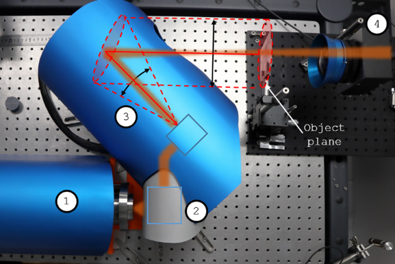
Full-field acquisition at 2.5 THz
The imaging unit combines a 5 mW quantum cascade laser with an I2S TZCam microbolometer camera containing 240 by 320 pixels. An aspherical silicon lens forms the image in transmission. The source is coherent, which creates a practical difficulty: stationary interference and speckle can impose patterns that are unrelated to the sample. Instead of accepting those patterns as fixed-background noise, the system uses galvanometric beam steering to move and homogenize the illumination during acquisition. On the 60 by 60 mm field used in the paper, the arrangement records at 25 frames per second and reaches a reported signal-to-noise ratio of up to 50 dB.
The sample is placed on a rotation stage and turned continuously at 25 degrees per second. A background image acquired without the object provides the reference for normalization, so each frame describes relative transmission rather than raw detector response. Arranging the normalized projections by viewing angle produces the sinogram from which the volume is reconstructed. This full-field strategy is the main reason acquisition is fast: each camera exposure captures an entire projection, whereas a raster system would have to visit every lateral position before advancing to the next angle.
Speed also reduces the number of angular samples available to reconstruction. The paper therefore uses a regularized three-dimensional implementation of the TerahertzTerahertz radiation is electromagnetic energy commonly associated with frequencies around 0.1 to 10 THz, between microwaves and infrared, where many materials reveal distinctive propagation, absorption, and imaging behavior. More Ordered Subsets Convex algorithm, or THz-OSC. It divides the projection data into subsets, updates a provisional estimate from each subset and combines those updates within a convex optimization framework. The method is intended to remain useful when the data contain fewer projections and more noise than conventional computed tomography would normally tolerate. The reconstructed voxel values retain a relationship to attenuation through the Beer-Lambert law, giving the volume a quantitative basis rather than treating it solely as a surface model.
From a reconstructed volume to a vascular skeleton
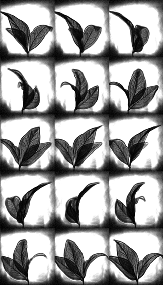
The test object consists of three intertwined leaves, chosen because their overlapping surfaces and branching venation create a demanding three-dimensional structure. The reconstruction preserves the general leaf geometry and makes the denser vascular network visible. K-means clustering separates voxel intensities into groups associated with background, leaf tissue and more strongly attenuating veins. Connected-component labeling then identifies coherent regions so that each leaf can be handled independently. These steps avoid relying on a single manually selected threshold, although the physical meaning of a cluster still depends on the contrast and noise of the acquired dataset.
A progressive thinning procedure reduces the segmented vascular volume to a one-voxel-wide centerline while attempting to preserve its topology. Nodes mark bifurcations and branches connect those nodes, producing a compact representation of an otherwise large volume. For this sample, the extracted skeleton contains 613 branches and 2,588 nodes. Its longest path includes 63 segments and 62 generations of branching. The authors use cross-sections along that path to estimate internal lumen diameter and vein-wall thickness in voxel units. The reported lumen varies between roughly 0.5 and 2.5 voxels, while wall-thickness measurements become more variable near the leaf extremities.
That variation illustrates both the analytical potential and the present limits. Once a skeleton has been established, the pipeline can search for local constrictions, changes in diameter or irregular branching without revisiting every voxel manually. At the same time, values close to a single voxel are sensitive to segmentation choices and spatial resolution. The reduced contrast near the leaf tips is associated with the camera’s depth of field, so apparent dimensional changes there cannot automatically be assigned to the biological structure. The measurements are therefore best read as morphology extracted under the stated imaging conditions, not as a universal metrology performance for every material and geometry.
Scope for rapid non-destructive inspection
The study demonstrates a complete laboratory workflow for fast terahertzTerahertz radiation is electromagnetic energy commonly associated with frequencies around 0.1 to 10 THz, between microwaves and infrared, where many materials reveal distinctive propagation, absorption, and imaging behavior. More tomography of a thin dielectric object: coherent illumination is averaged, full projections are collected during continuous rotation, a tailored iterative algorithm reconstructs attenuation, and automated processing converts the volume into structural descriptors. The non-ionizing measurement is attractive for samples that transmit at 2.5 THz, but it is not a replacement for X-ray tomography in all cases. Water-rich, strongly absorbing, metallic or thick objects may be inaccessible, and spatial resolution, depth of field and available source power continue to define what can be measured reliably.
Industrial transfer would require tests on the actual component families of interest, including repeatability, calibration against known dimensions, motion stability and an assessment of how acquisition speed affects defect-detection probability. Multispectral measurements could add material contrast, while improved optics could reduce the depth-of-field artifacts observed at the sample extremities. The collaboration represented in the paper brings together terahertzTerahertz radiation is electromagnetic energy commonly associated with frequencies around 0.1 to 10 THz, between microwaves and infrared, where many materials reveal distinctive propagation, absorption, and imaging behavior. More instrumentation, reconstruction and image-analysis expertise. Its most transferable contribution is the integration of those skills into one timed process, showing how terahertzTerahertz radiation is electromagnetic energy commonly associated with frequencies around 0.1 to 10 THz, between microwaves and infrared, where many materials reveal distinctive propagation, absorption, and imaging behavior. More volumes can become machine-readable inspection data rather than isolated research images.
Bibliographic reference
Recommended citation: Chopard, A., Guillet, J.-P., Gellie, P., Recur, B., Balacey, H., & Mounaix, P. (2023). Skeletonization and 3D rendering with real time terahertzTerahertz radiation is electromagnetic energy commonly associated with frequencies around 0.1 to 10 THz, between microwaves and infrared, where many materials reveal distinctive propagation, absorption, and imaging behavior. More tomography. Optics Continuum, 2(5), 1060. https://doi.org/10.1364/OPTCON.486227
Publisher: Optica Publishing Group. Airtable record: rec47n4mE9DoaZ5W4.