Blood plasma is a strongly hydrated and compositionally complex liquid, so its terahertzTerahertz radiation is electromagnetic energy commonly associated with frequencies around 0.1 to 10 THz, between microwaves and infrared, where many materials reveal distinctive propagation, absorption, and imaging behavior. More response reflects the combined behavior of water, proteins, lipids, ions and other dissolved constituents. This preclinical study asks whether experimentally induced tumor growth in mice is accompanied by measurable changes in that response. It combines reflection-mode terahertzTerahertz radiation is electromagnetic energy commonly associated with frequencies around 0.1 to 10 THz, between microwaves and infrared, where many materials reveal distinctive propagation, absorption, and imaging behavior. More time-domain spectroscopy with numerical electromagnetic modeling across 0.1-1.0 THz.
Featured visual: Image 1 from the Airtable record associated with this publication. Consult the original paper for the authoritative figure caption and interpretation. Source publication.
Visuals are drawn from the Airtable research archive. Figure numbering, rights and interpretation should be checked against the original publication before republication outside this site.
A controlled study of plasma in an animal tumor model
The experiment compared blood-related samples from control mice and mice grafted with Ehrlich carcinoma cells. The reported protocol involved nine control donors and ten animals receiving a subcutaneous injection of 0.2 mL of a suspension containing 106 cells per milliliter. Samples were collected on day 14. Whole blood was placed in K2EDTA tubes, and plasma was separated by centrifugation at 1,500 rpm for five minutes. A suspension of tumor cells in 0.9 percent saline was also examined.
These details define the scope of the evidence. The study concerns a specific mouse model, a modest number of animals and ex vivo liquid samples. It does not evaluate human screening, clinical diagnosis, treatment response or disease specificity. Differences between the control and grafted groups could reflect several biological changes associated with this model, and the paper does not establish a unique molecular cause for the measured terahertzTerahertz radiation is electromagnetic energy commonly associated with frequencies around 0.1 to 10 THz, between microwaves and infrared, where many materials reveal distinctive propagation, absorption, and imaging behavior. More contrast.
The use of plasma is nevertheless scientifically interesting. Whole blood contains cells whose dielectric response and volume fraction can dominate small biochemical variations. Plasma removes most of that cellular contribution while retaining a water-rich mixture of proteins, lipids, metabolites and ions. If tumor growth changes hydration, macromolecular concentration or particle-size distributions, those alterations may influence collective dielectric relaxation in the sub-terahertz band.
Reflection spectroscopy and numerical interpretation
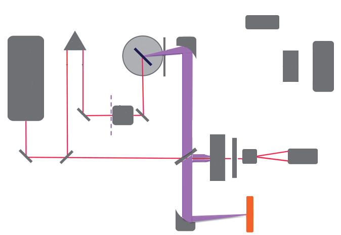
Broadband terahertzTerahertz radiation is electromagnetic energy commonly associated with frequencies around 0.1 to 10 THz, between microwaves and infrared, where many materials reveal distinctive propagation, absorption, and imaging behavior. More pulses were generated from undoped InAs excited by a femtosecond source operating at a wavelength of 1,040 nm, with 46 fs pulses at 70 MHz. Electro-optic detection used a CdTe crystal, balanced photodetection and lock-in amplification. For the reflection measurement, the liquid was held against a silicon wafer approximately 1.04 mm thick with a refractive index near 3.4. This geometry limited drying and provided a calibrated interface from which the sample-dependent reflection could be retrieved.
Fourier transformation of the temporal waveform yielded complex spectra. By comparing the sample response with the silicon reference and applying the appropriate interface relations, the team estimated refractive index, absorption coefficient and the real and imaginary parts of permittivity. The analysis focused on 0.1-1.0 THz, where signal quality and the dielectric response of hydrated matter were suitable for comparison.
The study also used finite-difference time-domain simulations. A numerical phantom represented a saline-filled volume containing an inhomogeneity with plasma-like properties. Simulated reflections at 0.7 and 0.8 THz reproduced the timing of the principal experimental interfaces and yielded reflection coefficients consistent with measurements over approximately 0.5-0.8 THz. Modeling did not identify a biochemical mechanism by itself; it tested whether a region with the measured dielectric contrast could account for the observed electromagnetic response.
Measured differences and their limits
Group differences were reported in several frequency intervals. Around 0.5-0.6 THz, the refractive index of whole blood was approximately 2.09 in controls and 2.00 in the tumor-bearing group. Plasma values were higher overall, near 2.20 and 2.10 respectively in the same broad comparison. Plasma from the grafted animals showed a 17-23 percent lower absorption coefficient in the 0.6-0.8 THz band. Differences were also reported for both components of complex permittivity, although their direction and magnitude depended on frequency and sample type.
The authors considered water fraction, suspended-particle size and changes in plasma constituents as possible contributors. They cited particle-size differences between groups and noted that replacing water with cells or macromolecular material can reduce absorption. Those explanations remain hypotheses rather than a demonstrated causal chain. Temperature, sample handling, thickness, sex differences between groups and biological variability are all relevant when interpreting subtle dielectric measurements.
A stronger follow-up design would measure those covariates directly, randomize the acquisition order and test whether a model trained on one animal cohort predicts a separately collected cohort. This would distinguish a stable electromagnetic effect from batch-specific variation.
For terahertzTerahertz radiation is electromagnetic energy commonly associated with frequencies around 0.1 to 10 THz, between microwaves and infrared, where many materials reveal distinctive propagation, absorption, and imaging behavior. More biophotonics, the important result is methodological: reflection THz-TDS detected reproducible group-level differences in a strongly absorbing liquid, and an electromagnetic simulation helped connect measured permittivity to reflected waveforms. The result supports continued investigation of biological-fluid spectroscopy and measurement phantoms. It does not show that the method can diagnose cancer, distinguish tumor types or replace established laboratory tests.
The author list reflects collaboration across terahertzTerahertz radiation is electromagnetic energy commonly associated with frequencies around 0.1 to 10 THz, between microwaves and infrared, where many materials reveal distinctive propagation, absorption, and imaging behavior. More instrumentation, numerical modeling and biological experimentation. Such collaboration is essential because a spectral difference has meaning only when the sample protocol, pathology model and electromagnetic retrieval are all controlled. Larger, balanced studies with blinded analysis, repeated measurements and independent validation would be required before assessing any translational relevance.
Publication details and citation
Recommended citation: Smolyanskaya, O. A., Kravtsenyuk, O. V., Panchenko, A. V., Odlyanitskiy, E. L., Guillet, J. P., Cherkasova, O. P., & Khodzitsky, M. K. (2017). Study of blood plasma optical properties in mice grafted with Ehrlich carcinoma in the frequency range 0.1-1.0 THz. Quantum Electronics, 47(11), 1031-1040. https://doi.org/10.1070/QEL16383
Record ID: recar92qjU7BJxLxV
Research themes: THz-TDS, blood plasma, complex permittivity, hydrated biological liquids, FDTD modeling, preclinical spectroscopy.