Near-field terahertzTerahertz radiation is electromagnetic energy commonly associated with frequencies around 0.1 to 10 THz, between microwaves and infrared, where many materials reveal distinctive propagation, absorption, and imaging behavior. More imaging seeks to overcome the diffraction limit by measuring the field extremely close to a sub-wavelength sensor. The NearSense project explored whether this approach could be implemented in silicon around 0.5 THz and eventually studied on freshly excised breast tissue. This publication reports progress on both sides of that problem: identifying a useful frequency window from ex vivo measurements and developing an integrated SiGe sensor capable of resolving small test structures.
Featured visual: Contextual research figure from âTerahertzTerahertz radiation is electromagnetic energy commonly associated with frequencies around 0.1 to 10 THz, between microwaves and infrared, where many materials reveal distinctive propagation, absorption, and imaging behavior. More refractive index-based morphological dilation for breast carcinoma delineationâ. It illustrates a closely related terahertzTerahertz radiation is electromagnetic energy commonly associated with frequencies around 0.1 to 10 THz, between microwaves and infrared, where many materials reveal distinctive propagation, absorption, and imaging behavior. More topic and is not a figure from the publication discussed on this page. Source publication.

Visuals are drawn from the Airtable research archive. Figure numbering, rights and interpretation should be checked against the original publication before republication outside this site.
Connecting tissue contrast to a silicon frequency band
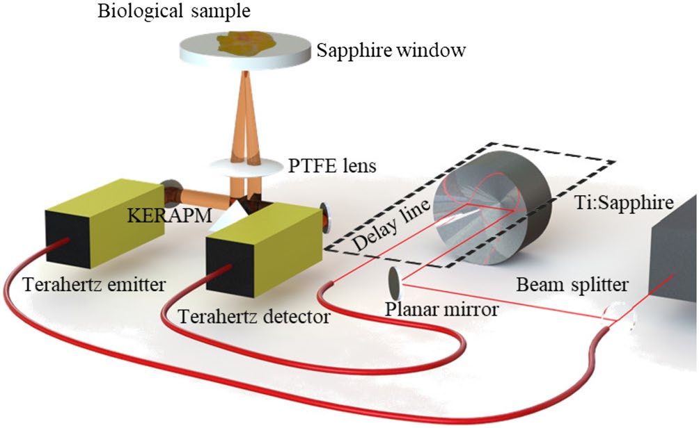
TerahertzTerahertz radiation is electromagnetic energy commonly associated with frequencies around 0.1 to 10 THz, between microwaves and infrared, where many materials reveal distinctive propagation, absorption, and imaging behavior. More measurements of biological tissue are dominated by hydration, dielectric composition and interfaces. Frequencies in the 300-600 GHz region offer a compromise: they can provide contrast among tissue compositions while remaining accessible to advanced silicon-germanium electronics. The project examined that band with time-domain measurements on 17 freshly excised breast-tissue samples representing several pathological and non-pathological tissue compositions.
The broadband measurements indicated detectable differences between adipose tissue, fibrous tissue and regions containing malignant and fibrous components. Lower parts of the selected band offered stronger dielectric contrast in some comparisons, while operation toward 500-600 GHz provided a smaller free-space wavelength and therefore better conventional spatial resolution. Those results guided the design frequency of the electronic sensor rather than establishing a diagnostic threshold.
This distinction is essential. The tissue work was ex vivo, used a limited sample collection and measured composite regions whose water, fat and fiber content differed. It did not demonstrate clinical sensitivity or specificity, prospective margin detection, patient outcomes or use during surgery. The publication presents a technological research direction based on measurable dielectric contrast.
A sub-wavelength SiGe near-field sensor
The electronic device was fabricated in a 0.13 micrometer silicon-germanium heterojunction bipolar transistor process. It operated over approximately 534-562 GHz and incorporated a resonant near-field structure designed to confine the interaction to a region much smaller than the free-space wavelength. In this regime, lateral resolution is determined by the size and spacing of the transducer and sample, not solely by far-field focusing.
Low-frequency and oscillator noise are serious problems when the sensor responds to small permittivity changes. The team modulated, or chopped, the oscillator and used synchronous detection to move the measurement away from the strongest flicker-noise region. The paper reports a substantial signal-to-noise improvement, from a quoted 42 dB in direct operation to as much as 115 dB under the selected chopping conditions. That figure belongs to the reported electronic test configuration and should not be interpreted as tissue-classification performance.
The sensor was mounted in a modified scanning platform with controlled vertical position and a high-precision lateral piezo stage. Nickel mesh provided a known imaging target. One mesh had bars 50 micrometers wide with a 250 micrometer pitch. Scans with 10 micrometer lateral steps reproduced the periodic structure and measured a bar width near 60 micrometers, the difference being consistent with sampling and system geometry. The underlying near-field design was associated with a nominal lateral interaction scale around 10-12 micrometers, although the demonstrated image and acquisition steps did not constitute a biological image at that resolution.
The scans also remained slow, with integration times of several seconds per pixel in the reported tests. Moving from a single experimental sensor to a practical imaging tool would require arrays, controlled sample contact or spacing, robust calibration and much shorter acquisition times. Fresh tissue introduces surface relief, drying and motion that a flat metal mesh does not reproduce.
What NearSense establishes
The work establishes compatibility between three elements: a biologically relevant sub-terahertz band, a silicon process capable of generating and detecting signals in that band, and a near-field structure that images sub-wavelength features on a controlled target. This is an important integration result because compact electronic arrays could eventually be more scalable than a free-space ultrafast-laser system.
It does not establish a medical device. Before any clinical role could be assessed, later studies would need larger and representative tissue cohorts, blinded comparison with histopathology, pre-defined performance metrics, reproducible handling protocols and safety and workflow evaluation. Near-field measurements would also need a well-controlled relationship between the sensor and a moist, uneven specimen.
The project is intrinsically collaborative. The author list joins high-frequency silicon design, terahertzTerahertz radiation is electromagnetic energy commonly associated with frequencies around 0.1 to 10 THz, between microwaves and infrared, where many materials reveal distinctive propagation, absorption, and imaging behavior. More spectroscopy, biomedicalTerahertz and millimeter-wave technologies offer promising non-ionizing tools for biomedical tissue analysis, particularly for breast cancer research. Their sensitivity to water content, tissue structure, and dielectric contrast can help distinguish... More sample work and pathology expertise. That breadth is appropriate because neither a promising tissue spectrum nor an advanced chip is sufficient on its own. The publication records progress toward connecting them, while leaving validation and translation as future research.
Equally important, an array would need reference pixels and repeatable distance control so that variations in chip gain or sample topography are not confused with dielectric contrast. Those engineering controls are prerequisites for meaningful biological comparison.
For the wider terahertzTerahertz radiation is electromagnetic energy commonly associated with frequencies around 0.1 to 10 THz, between microwaves and infrared, where many materials reveal distinctive propagation, absorption, and imaging behavior. More field, NearSense illustrates how application requirements can shape component design. The target contrast selected the frequency range; the desired resolution motivated near-field confinement; and the small signal drove modulation and noise control. The result is best understood as a platform advance for ex vivo research, not as evidence of clinical identification of breast tumors.
Publication details and citation
Recommended citation: Mavarani, L., Hillger, P., Bucher, T., Grzyb, J., Pfeiffer, U. R., Cassar, Q., Al-Ibadi, A., Zimmer, T., Guillet, J.-P., Mounaix, P., & MacGrogan, G. (2018). NearSense – Advances Towards a Silicon-Based TerahertzTerahertz radiation is electromagnetic energy commonly associated with frequencies around 0.1 to 10 THz, between microwaves and infrared, where many materials reveal distinctive propagation, absorption, and imaging behavior. More Near-Field Imaging Sensor for Ex Vivo Breast Tumour Identification. Frequenz, 72(3-4), 93-99. https://doi.org/10.1515/freq-2018-0016
Record ID: recpQme9iaQ1FLJD5
Research themes: near-field terahertzTerahertz radiation is electromagnetic energy commonly associated with frequencies around 0.1 to 10 THz, between microwaves and infrared, where many materials reveal distinctive propagation, absorption, and imaging behavior. More imaging, SiGe electronics, 500-600 GHz sensors, ex vivo tissue research, sub-wavelength imaging.