Research review · Multimodal biomedicalTerahertz and millimeter-wave technologies offer promising non-ionizing tools for biomedical tissue analysis, particularly for breast cancer research. Their sensitivity to water content, tissue structure, and dielectric contrast can help distinguish... More optics
Multimodal Optical Diagnostics of Glycated Biological Tissues
Persistent hyperglycemia changes biological matter through non-enzymatic glycation. Glucose first reacts with amino groups in proteins, then forms more stable products and, over time, advanced glycation end-products that can alter molecular structure, hydration, scattering and refractive index. This review asks how optical measurements, from visible wavelengths to the terahertzTerahertz radiation is electromagnetic energy commonly associated with frequencies around 0.1 to 10 THz, between microwaves and infrared, where many materials reveal distinctive propagation, absorption, and imaging behavior. More range, can observe those changes. Rather than presenting one diagnostic instrument, it compares a family of complementary techniques and argues that phase, spectral and structural information should be interpreted together. The article is therefore a map of physical contrasts associated with glycation, not evidence that any single optical signal can diagnose diabetes or its complications.
Conventional glucose measurements and glycated hemoglobin assays answer different clinical questions: one reflects a momentary concentration, while HbA1c is used as an indicator of longer-term glycemic exposure. Optical research seeks additional information from blood, plasma, cells and tissue without relying solely on chemical separation. The challenge is that glycation does not create one isolated, universally measurable signature. It modifies proteins in an aqueous, heterogeneous environment where temperature, concentration, tissue architecture and hydration can all affect the same observable. A useful optical protocol must therefore separate a small biochemical effect from these confounding variables and be validated against accepted laboratory measurements.
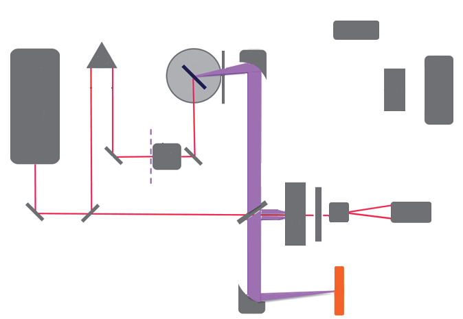
Featured visual: Contextual research figure from âStudy of blood plasma optical properties in mice grafted with Ehrlich carcinoma in the frequency range 0.1â1.0 THzâ. It illustrates a closely related terahertzTerahertz radiation is electromagnetic energy commonly associated with frequencies around 0.1 to 10 THz, between microwaves and infrared, where many materials reveal distinctive propagation, absorption, and imaging behavior. More topic and is not a figure from the publication discussed on this page. Source publication.
Visuals are drawn from the Airtable research archive. Figure numbering, rights and interpretation should be checked against the original publication before republication outside this site.
Connecting phase, spectroscopy and tissue structure
The review begins with refractometry and phase-sensitive microscopy because glycation can change the complex refractive index of protein solutions. In one set of measurements discussed by the authors, oxygenated and glycated hemoglobin solutions at 3 g L-1 were examined between 480 and 1550 nm. The reported refractive-index difference between the two forms ranged from approximately 0.001 to 0.006 across that interval. Temperature also changed the measured index, with an approximately linear trend for both solutions. These results illustrate both the attraction and the difficulty of phase diagnostics: a measurable contrast exists, but its interpretation requires stable concentration, thermal control and a reference composition.
Digital holographic microscopy and diffraction tomography extend this idea from a bulk reading to spatially resolved phase. They infer optical path length from the phase delay introduced by a sample, allowing researchers to investigate cells or distributions of material rather than only an average value. Optical coherence tomography adds depth-resolved scattering information, while optical coherence microscopy can examine smaller fields at higher resolution. None of these measurements is chemically specific by itself. Changes in cell shape, aggregation, protein concentration or surrounding medium can all shift the recorded signal. Their value grows when phase information is paired with a modality that responds to a different physical property.
The authors consequently discuss fluorescence, Raman methods and coherent anti-Stokes Raman scattering as complementary sources of molecular contrast. Fluorescence can be sensitive to advanced glycation end-products, while vibrational spectroscopy probes molecular bonds and composition. A combined system can ask whether a phase change and a molecular marker occur in the same region, reducing reliance on a single indirect indicator. This is the central meaning of “multimodal” in the review: not simply placing several instruments together, but using independent observables to constrain an interpretation that would otherwise remain ambiguous.
Optical clearing provides another experimental lens on glycated tissue. Clearing agents reduce scattering by changing refractive-index matching and water distribution within a sample. Monitoring how such an agent diffuses can reveal differences in permeability, hydration and extracellular structure. The reviewed work includes diabetic animal models and measurements of microhemodynamics, but these remain model-based investigations of tissue physiology. They should not be read as a validated human screening protocol. Agent concentration, diffusion time, tissue preparation and vascular state are all part of the measured response.
TerahertzTerahertz radiation is electromagnetic energy commonly associated with frequencies around 0.1 to 10 THz, between microwaves and infrared, where many materials reveal distinctive propagation, absorption, and imaging behavior. More spectroscopy contributes sensitivity to low-frequency collective motions and to water. That sensitivity makes it relevant to glycation research because protein conformation and hydration are intertwined, but it also creates a strong confounder: a change in water content can dominate a subtle molecular contribution. The review considers spectral analysis methods such as moving-window partial least squares for extracting concentration-related information from complex data. It also discusses using Kramers-Kronig relationships to connect absorption and refractive-index behavior. These tools can improve quantitative analysis, yet their predictions remain dependent on calibration sets and on whether future samples resemble the material used to build the model.
What a multimodal strategy can establish
Taken together, the studies surveyed in the article show that glycated and non-glycated materials can produce distinguishable optical responses under controlled conditions. Refractive index, phase delay, scattering, fluorescence, Raman bands and terahertzTerahertz radiation is electromagnetic energy commonly associated with frequencies around 0.1 to 10 THz, between microwaves and infrared, where many materials reveal distinctive propagation, absorption, and imaging behavior. More dielectric properties each expose a different part of the system. The evidence is strongest when the sample type is defined, composition is controlled and the optical result is compared with a biochemical reference. It becomes weaker when a contrast observed in a solution or animal model is generalized directly to intact human tissue.
The review also makes clear why a single “glycation image” is unlikely to be sufficient. Biological tissues contain several proteins, lipids, cells and extracellular structures, all affected differently by disease and sample handling. Water strongly influences both near-infrared and terahertzTerahertz radiation is electromagnetic energy commonly associated with frequencies around 0.1 to 10 THz, between microwaves and infrared, where many materials reveal distinctive propagation, absorption, and imaging behavior. More signals. Optical clearing intentionally changes hydration and scattering. Temperature affects refractive index, and fluorescence may depend on the specific advanced glycation products present. A future diagnostic study would therefore need prospective cohorts, standardized preparation, independent HbA1c or biochemical measurements, and statistical testing on samples not used for model development.
The broad collaboration behind the review brings together photonics, terahertzTerahertz radiation is electromagnetic energy commonly associated with frequencies around 0.1 to 10 THz, between microwaves and infrared, where many materials reveal distinctive propagation, absorption, and imaging behavior. More spectroscopy, biophysics and medical research. That range is appropriate to the problem: instrument specialists can measure weak phase or spectral variations, while biological and clinical partners define meaningful samples and reference standards. The article supports continued collaboration around calibrated multimodal experiments, especially where two or more observables can be acquired from the same specimen. It does not establish a replacement for blood glucose or HbA1c testing, nor does it demonstrate a medical device ready for patient care.
Its durable contribution is a framework for asking better questions. Instead of treating glycation as a single optical target, the review connects molecular chemistry to physical properties and then to measurement design. That framework can guide studies of plasma, hemoglobin, cells, skin or model tissues, provided each study states what was measured and what remained inferred. TerahertzTerahertz radiation is electromagnetic energy commonly associated with frequencies around 0.1 to 10 THz, between microwaves and infrared, where many materials reveal distinctive propagation, absorption, and imaging behavior. More methods occupy a useful place in this spectrum because they probe hydration and collective dynamics, but their results gain credibility when combined with phase imaging, vibrational spectroscopy and rigorous biochemical controls.
Publication and citation
Recommended citation: Smolyanskaya, O. A., Lazareva, E. N., Nalegaev, S. S., Petrov, N. V., Zaytsev, K. I., Timoshina, P. A., Tuchina, D. K., Toropova, Ya. G., Kornyushin, O. V., Babenko, A. Yu., Guillet, J.-P., & Tuchin, V. V. (2019). Multimodal optical diagnostics of glycated biological tissues. Biochemistry (Moscow), 84(S1), 124-143. https://doi.org/10.1134/S0006297919140086
Publisher: Pleiades Publishing Ltd. Airtable record: recspxrNhWNLAv0P9.