TerahertzTerahertz radiation is electromagnetic energy commonly associated with frequencies around 0.1 to 10 THz, between microwaves and infrared, where many materials reveal distinctive propagation, absorption, and imaging behavior. More biophotonics is shaped by a paradox: water makes biological matter strongly absorbing, yet the dielectric dynamics of water and hydration shells are also a major source of useful contrast. This extensive review organizes the field around that tension. It connects physical models of aqueous media with spectroscopy, imaging, near-field methods, waveguides, tissue phantoms and exploratory biomedicalTerahertz and millimeter-wave technologies offer promising non-ionizing tools for biomedical tissue analysis, particularly for breast cancer research. Their sensitivity to water content, tissue structure, and dielectric contrast can help distinguish... More applications.
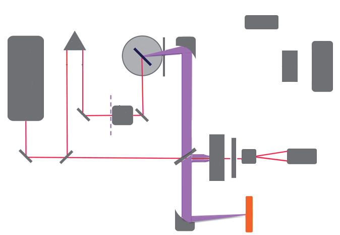
Featured visual: Contextual research figure from âStudy of blood plasma optical properties in mice grafted with Ehrlich carcinoma in the frequency range 0.1â1.0 THzâ. It illustrates a closely related terahertzTerahertz radiation is electromagnetic energy commonly associated with frequencies around 0.1 to 10 THz, between microwaves and infrared, where many materials reveal distinctive propagation, absorption, and imaging behavior. More topic and is not a figure from the publication discussed on this page. Source publication.

Visuals are drawn from the Airtable research archive. Figure numbering, rights and interpretation should be checked against the original publication before republication outside this site.
Water as signal, background and limiting medium
In the terahertzTerahertz radiation is electromagnetic energy commonly associated with frequencies around 0.1 to 10 THz, between microwaves and infrared, where many materials reveal distinctive propagation, absorption, and imaging behavior. More range, liquid water exhibits strong dielectric relaxation associated with collective molecular motion and hydrogen-bond dynamics. Biological tissues contain bulk-like water, water constrained near membranes and macromolecules, dissolved ions and structural solids. Their measured permittivity therefore depends on water fraction, binding state, temperature and composition, not on a single isolated molecular resonance.
The review describes dielectric models built from Debye-type relaxation terms, damped oscillators, high-frequency permittivity and ionic conductivity. Such models represent both the real part of permittivity, which governs phase velocity and reflection, and the imaginary part, which is associated with loss. Effective-medium formulations extend the description to mixtures in which cells, proteins or inclusions occupy part of an aqueous host.
This modeling is more than a theoretical exercise. Extracting refractive index or absorption from a time-domain waveform requires an interface model, known sample thickness and stable reference. Hydrated samples can dry, warm or change shape during acquisition. A difference between tissues may arise from water content or surface contact rather than a disease-specific molecular feature. The review repeatedly shows why controlled phantoms and careful sample protocols are necessary.
Measurement and imaging strategies
TerahertzTerahertz radiation is electromagnetic energy commonly associated with frequencies around 0.1 to 10 THz, between microwaves and infrared, where many materials reveal distinctive propagation, absorption, and imaging behavior. More time-domain spectroscopy is central because it records the electric field as a function of time and retrieves amplitude and phase over a broad band after Fourier transformation. Frequency-domain, Fourier-transform and high-resolution electronic methods provide complementary combinations of bandwidth, spectral resolution and acquisition speed. Reflection geometry is often necessary for thick or highly absorbing tissues, while transmission can be effective for thin sections, liquids in calibrated cells and low-loss samples.
The review surveys photoconductive antennas, optical rectification, electronic sources, electro-optic sampling and several detector families. It also considers waveguides and probe geometries intended to bring radiation to constrained locations. Near-field techniques, apertures, resonant structures and metamaterial concepts can produce spatial detail below the free-space diffraction limit, although their working distance and sample presentation are demanding.
Strong water absorption limits penetration, prompting research on dehydration, hyperosmotic agents and optical clearing. These interventions may increase measurable depth, but they also alter the tissue being studied and must be treated as experimental manipulations rather than neutral improvements. Thermal models are likewise important because absorbed terahertzTerahertz radiation is electromagnetic energy commonly associated with frequencies around 0.1 to 10 THz, between microwaves and infrared, where many materials reveal distinctive propagation, absorption, and imaging behavior. More power can raise local temperature. Exposure conditions, duty cycle and heat transport need to be quantified whenever biological effects or repeated measurements are considered.
Multispectral and multitemporal processing can combine frequency, phase and spatial information. Pattern recognition and machine learning may help separate broad, overlapping responses, but model performance depends on cohort design, preprocessing and independent validation. For aqueous biological materials, the review does not support a simplistic catalog of sharp spectral fingerprints analogous to gas-phase spectroscopy.
Biomedical questions and appropriate caution
The article surveys studies of skin, breast and oral tissues, blood-related liquids, corneal hydration and tear-film dynamics. Reported contrasts are often linked to differences in water content, cell density, fibrosis, necrosis or bound-water fractions. TerahertzTerahertz radiation is electromagnetic energy commonly associated with frequencies around 0.1 to 10 THz, between microwaves and infrared, where many materials reveal distinctive propagation, absorption, and imaging behavior. More reflectometry can be especially sensitive to superficial hydration, while sectioned or excised tissues can be mapped against histology under controlled laboratory conditions.
These examples establish a research field, not a portfolio of validated clinical tools. Results obtained from phantoms, cultured samples, animal models or freshly excised human tissue cannot be generalized directly to in vivo diagnosis. Many studies are small, use different instruments and sample preparations, and report contrast rather than prospective clinical accuracy. The review’s value is to expose those technical dependencies and identify what must be standardized.
Tissue-equivalent phantoms are one response to the reproducibility problem. By controlling water, polymer, protein or particulate content, researchers can create targets with known geometry and approximate dielectric properties. Phantoms support calibration, inter-instrument comparison, resolution testing and thermal safety experiments without biological variability. They cannot reproduce every aspect of living tissue, but they provide a necessary bridge between instrument development and biomedicalTerahertz and millimeter-wave technologies offer promising non-ionizing tools for biomedical tissue analysis, particularly for breast cancer research. Their sensitivity to water content, tissue structure, and dielectric contrast can help distinguish... More studies.
Ocular applications illustrate both opportunity and difficulty. The cornea and tear film are superficial and hydration-sensitive, making reflection measurements physically plausible. At the same time, motion, curvature, safety limits and the very small thickness of the tear film place stringent requirements on range resolution and calibration. Similar application-specific constraints recur throughout the review.
A collaborative map of the field
With authors spanning terahertzTerahertz radiation is electromagnetic energy commonly associated with frequencies around 0.1 to 10 THz, between microwaves and infrared, where many materials reveal distinctive propagation, absorption, and imaging behavior. More physics, spectroscopy, imaging and biophotonics, the paper synthesizes a broad international research landscape. Its 77 pages connect fundamental dielectric theory to hardware and biological experiments. This breadth is useful for identifying shared problems: water-dominated loss, inconsistent sample protocols, limited penetration, the need for sub-wavelength resolution and the risk of over-interpreting statistical separation.
The review suggests that progress will depend less on a single ideal frequency than on matched systems. Source, detector, geometry, sample preparation, physical model and analysis must be chosen together for a defined question. High-resolution spectroscopy can examine liquids and biomolecular systems; reflection imaging can map superficial tissue; near-field sensors can interrogate small ex vivo regions; and phantoms can make performance claims testable.
No clinical claim follows from the review itself. It provides scientific rationale and a record of experimental approaches available by 2018. Translation would require standardized acquisition, safety evaluation, representative human cohorts, blinded comparison with accepted references and evidence that a terahertzTerahertz radiation is electromagnetic energy commonly associated with frequencies around 0.1 to 10 THz, between microwaves and infrared, where many materials reveal distinctive propagation, absorption, and imaging behavior. More measurement improves a real decision. Within those limits, the article remains a substantial guide to why biological matter is challenging in the terahertzTerahertz radiation is electromagnetic energy commonly associated with frequencies around 0.1 to 10 THz, between microwaves and infrared, where many materials reveal distinctive propagation, absorption, and imaging behavior. More band and why that challenge continues to generate valuable physics and instrumentation research.
Recommended citation: Smolyanskaya, O. A., Chernomyrdin, N. V., Konovko, A. A., Zaytsev, K. I., Ozheredov, I. A., Cherkasova, O. P., Nazarov, M. M., Guillet, J.-P., Kozlov, S. A., Kistenev, Yu. V., Coutaz, J.-L., Mounaix, P., Vaks, V. L., Son, J.-H., Cheon, H., Wallace, V. P., Feldman, Yu., Popov, I., Yaroslavsky, A. N., Shkurinov, A. P., & Tuchin, V. V. (2018). TerahertzTerahertz radiation is electromagnetic energy commonly associated with frequencies around 0.1 to 10 THz, between microwaves and infrared, where many materials reveal distinctive propagation, absorption, and imaging behavior. More biophotonics as a tool for studies of dielectric and spectral properties of biological tissues and liquids. Progress in Quantum Electronics, 62, 1-77. https://doi.org/10.1016/j.pquantelec.2018.10.001
Record ID: rectcnyu1ADB4oPdf
Research themes: terahertzTerahertz radiation is electromagnetic energy commonly associated with frequencies around 0.1 to 10 THz, between microwaves and infrared, where many materials reveal distinctive propagation, absorption, and imaging behavior. More biophotonics, dielectric spectroscopy, water relaxation, tissue imaging, near-field methods, biological phantoms.