Research publication · TerahertzTerahertz radiation is electromagnetic energy commonly associated with frequencies around 0.1 to 10 THz, between microwaves and infrared, where many materials reveal distinctive propagation, absorption, and imaging behavior. More plasma-pellet holography
Fast TerahertzTerahertz radiation is electromagnetic energy commonly associated with frequencies around 0.1 to 10 THz, between microwaves and infrared, where many materials reveal distinctive propagation, absorption, and imaging behavior. More Spectroscopic Holographic Assessment of Optical Properties of Diabetic Blood Plasma
Liquid blood plasma is dominated by water in the terahertzTerahertz radiation is electromagnetic energy commonly associated with frequencies around 0.1 to 10 THz, between microwaves and infrared, where many materials reveal distinctive propagation, absorption, and imaging behavior. More range, making transmission difficult and leaving measurements sensitive to evaporation and handling time. This study instead freeze-dries plasma and presses the resulting material into small pellets. TerahertzTerahertz radiation is electromagnetic energy commonly associated with frequencies around 0.1 to 10 THz, between microwaves and infrared, where many materials reveal distinctive propagation, absorption, and imaging behavior. More time-domain spectroscopy is used to measure their refractive index and absorption between 0.2 and 1.2 THz. Those experimental data then drive a numerical model of pulse time-domain holography, which tests whether a spatial map of optical properties could be reconstructed without point-by-point raster scanning. The reported comparison is exploratory and based on material from one diabetic and one non-diabetic source; it does not establish a clinical diabetes test.
The approach separates two questions that are sometimes conflated. The spectroscopy asks whether prepared pellets show different optical behavior. The holographic simulation asks whether a future full-field instrument could recover that behavior spatially from propagated terahertzTerahertz radiation is electromagnetic energy commonly associated with frequencies around 0.1 to 10 THz, between microwaves and infrared, where many materials reveal distinctive propagation, absorption, and imaging behavior. More fields. The second part is not a clinical measurement of a cohort and not a direct experimental demonstration of a finished camera. Its role is to connect measured material properties to an acquisition and reconstruction concept.
Featured visual: Image 1 from the Airtable record associated with this publication. Consult the original paper for the authoritative figure caption and interpretation. Source publication.
Visuals are drawn from the Airtable research archive. Figure numbering, rights and interpretation should be checked against the original publication before republication outside this site.
From lyophilized plasma to a simulated hologram
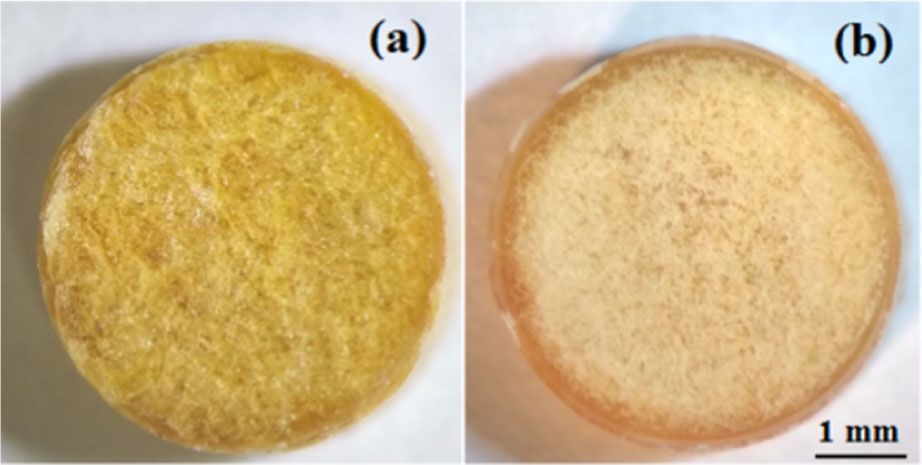
Plasma was freeze-dried into a porous solid containing proteins, triglycerides, fibrinogen and other nonvolatile constituents, then ground and pressed into pellets approximately 5 mm in diameter and 1.8 mm thick. Ten pellets were prepared for each of the two source groups described in the study. Lyophilization reduced water content and produced samples that were easier to store and transmit through than liquid plasma, but it also changed the physical state of the specimen. The experiment therefore measures a prepared plasma-derived material, not circulating blood in its native aqueous condition.
A commercial time-domain spectrometer generated terahertzTerahertz radiation is electromagnetic energy commonly associated with frequencies around 0.1 to 10 THz, between microwaves and infrared, where many materials reveal distinctive propagation, absorption, and imaging behavior. More pulses from an InAs emitter driven by femtosecond Ti:sapphire laser light. After transmission through a pellet, the electric field was sampled electro-optically in ZnTe. Fourier transformation supplied both amplitude and phase as functions of frequency. Measurements were made at nine spatial points on each pellet, allowing the authors to examine heterogeneity rather than treating every pressed sample as perfectly uniform.
A thin-slab transmission model converted the complex transmission coefficient into refractive index and extinction behavior, from which absorption could be derived. Pellet thickness and phase offset were part of the retrieval, and a linear extrapolation near zero frequency was used to correct the phase baseline. These processing choices matter because a small thickness error can appear as a refractive-index shift. Surface roughness and density variations introduced during pressing can likewise produce spatial changes that are not solely biochemical.
The holography branch began with a modeled single-cycle terahertzTerahertz radiation is electromagnetic energy commonly associated with frequencies around 0.1 to 10 THz, between microwaves and infrared, where many materials reveal distinctive propagation, absorption, and imaging behavior. More pulse. At each frequency, that field was multiplied by a spatially varying complex transmission function built from the experimentally retrieved pellet properties and thickness. An angular-spectrum calculation propagated the field 5 mm from the pellet to a detector plane. The simulation included a finite electro-optic response and added noise to approximate a physical acquisition. An inverse time transform then yielded a complete space-time field resembling what a pulse time-domain holographic detector could record.
Reconstruction numerically propagated the simulated detector field back toward the object plane. Dividing by the known incident spectrum recovered local transmission amplitude and phase, which were again converted to optical properties through the slab model. In principle, a large-area electro-optic detector and camera could record many spatial positions together, replacing a two-dimensional mechanical scan with a sequence over pulse delay. The study tests that reconstruction numerically with properties grounded in spectroscopy; it does not report the full experimental camera operating on these pellets.
Observed contrast and the limits of interpretation
The measured and reconstructed maps showed that the pellet prepared from diabetic plasma was more spatially heterogeneous and generally had higher refractive-index regions across the investigated band than the non-diabetic comparison. The authors relate this behavior to differences in glycated proteins and lipid composition. That interpretation is plausible within the biochemical context, but the experiment cannot isolate glycation as the sole cause. Pellet density, particle packing, remaining moisture, surface shape and individual donor composition can also affect the terahertzTerahertz radiation is electromagnetic energy commonly associated with frequencies around 0.1 to 10 THz, between microwaves and infrared, where many materials reveal distinctive propagation, absorption, and imaging behavior. More response.
The very small source population is the dominant evidence limit. Multiple pellets from one specimen provide information about preparation repeatability and within-sample variation; they do not substitute for independent participants. No diagnostic sensitivity, specificity or population-level separation can be inferred. Fasting glucose status and diabetes classification describe the two sources, while direct biochemical assays of each optical feature would be needed to establish which constituent produced the contrast.
Lyophilization is both the enabling step and a boundary on translation. Stable, transportable pellets could support inter-laboratory comparisons and reduce the overwhelming absorption of liquid water. Yet any future clinical workflow would include blood collection, plasma separation, freeze-drying, grinding and pressing. The effects of those steps on reproducibility, turnaround time and operator variability must be assessed against standard biochemical tests. Calling the optical reading label-free does not make the preparation immediate or non-invasive.
The international author team combines sample preparation and biomedicalTerahertz and millimeter-wave technologies offer promising non-ionizing tools for biomedical tissue analysis, particularly for breast cancer research. Their sensitivity to water content, tissue structure, and dielectric contrast can help distinguish... More optics with terahertzTerahertz radiation is electromagnetic energy commonly associated with frequencies around 0.1 to 10 THz, between microwaves and infrared, where many materials reveal distinctive propagation, absorption, and imaging behavior. More spectroscopy and computational holography. A robust next study would recruit an independent and diverse cohort, measure HbA1c and relevant plasma chemistry, randomize sample processing, and test a locked reconstruction model without knowledge of clinical group. Parallel work could build and validate the full-field holographic detector, comparing its maps directly with conventional raster-scanned TDS.
Within those boundaries, the paper offers a coherent proof of method. It shows that freeze-dried plasma pellets can transmit measurable terahertzTerahertz radiation is electromagnetic energy commonly associated with frequencies around 0.1 to 10 THz, between microwaves and infrared, where many materials reveal distinctive propagation, absorption, and imaging behavior. More pulses, that spatially varying complex optical properties can be retrieved, and that those measured properties support a simulated holographic reconstruction. The result is a platform for investigating plasma composition and faster field acquisition. It is not evidence that terahertzTerahertz radiation is electromagnetic energy commonly associated with frequencies around 0.1 to 10 THz, between microwaves and infrared, where many materials reveal distinctive propagation, absorption, and imaging behavior. More holography diagnoses diabetes, replaces glucose monitoring or is ready for point-of-care use.
Publication and citation
Recommended citation: Kulya, M. S., Odlyanitskiy, E. L., Cassar, Q., Mustafin, I. A., Trukhin, V. N., Gavrilova, P. G., Korolev, D. V., Kononova, Y. A., Balbekin, N. S., Mounaix, P., Guillet, J.-P., Petrov, N. V., & Smolyanskaya, O. A. (2020). Fast terahertzTerahertz radiation is electromagnetic energy commonly associated with frequencies around 0.1 to 10 THz, between microwaves and infrared, where many materials reveal distinctive propagation, absorption, and imaging behavior. More spectroscopic holographic assessment of optical properties of diabetic blood plasma. Journal of Infrared, Millimeter, and TerahertzTerahertz radiation is electromagnetic energy commonly associated with frequencies around 0.1 to 10 THz, between microwaves and infrared, where many materials reveal distinctive propagation, absorption, and imaging behavior. More Waves, 41(9), 1041-1056. https://doi.org/10.1007/s10762-020-00728-9
Publisher: Springer Science and Business Media LLC. Airtable record: recOl4qmCiFsxJIMM.