Research publication · Real-time terahertzTerahertz radiation is electromagnetic energy commonly associated with frequencies around 0.1 to 10 THz, between microwaves and infrared, where many materials reveal distinctive propagation, absorption, and imaging behavior. More 3D imaging
Shape-from-focus for real-time terahertzTerahertz radiation is electromagnetic energy commonly associated with frequencies around 0.1 to 10 THz, between microwaves and infrared, where many materials reveal distinctive propagation, absorption, and imaging behavior. More 3D imaging
Three-dimensional terahertzTerahertz radiation is electromagnetic energy commonly associated with frequencies around 0.1 to 10 THz, between microwaves and infrared, where many materials reveal distinctive propagation, absorption, and imaging behavior. More imaging is often associated with computed tomography, but tomography is not always the natural choice for a thin, bent or layered object. This study adapts shape-from-focus, a method developed in optical microscopy, to a real-time terahertzTerahertz radiation is electromagnetic energy commonly associated with frequencies around 0.1 to 10 THz, between microwaves and infrared, where many materials reveal distinctive propagation, absorption, and imaging behavior. More camera. The principle is direct: as a sample moves through the limited depth of field of an imaging system, different surface regions become sharp at different axial positions. Measuring that focus variation pixel by pixel produces both an all-in-focus image and a depth map. The authors demonstrate the approach on dried leaves, using fast full-field acquisition rather than a mechanically raster-scanned single detector.
The method addresses a limitation that is normally treated as an optical defect. A terahertzTerahertz radiation is electromagnetic energy commonly associated with frequencies around 0.1 to 10 THz, between microwaves and infrared, where many materials reveal distinctive propagation, absorption, and imaging behavior. More objective cannot keep an extended three-dimensional object perfectly focused when its depth exceeds the system’s depth of field. Shape-from-focus turns that blur into information. It is especially suited to surfaces or separated thin layers that transmit enough radiation to appear in the image stack. It does not reconstruct a general volumetric distribution in the way a tomographic inversion can; instead, it estimates the axial position at which visible structure is best focused.
Featured visual: Image 1 from the Airtable record associated with this publication. Consult the original paper for the authoritative figure caption and interpretation. Source publication.
Visuals are drawn from the Airtable research archive. Figure numbering, rights and interpretation should be checked against the original publication before republication outside this site.
Following focus through a rapid axial scan
The experimental platform used a quantum cascade laser operating at 2.5 THz and cooled to 44 K. Its maximum reported output power was 3 mW. A high-resistivity silicon hemispherical lens improved coupling and beam quality, while a quarter-magnification objective formed a 64 by 48 mm field of view on a microbolometer focal-plane array. The optical depth of field was approximately 6 mm, corresponding to a Rayleigh distance near 3 mm. This value ultimately sets the scale at which two axial surfaces can be distinguished.
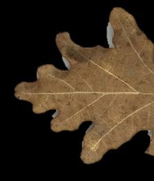
During acquisition, the sample translated along the optical axis at 1.25 mm s-1, and the camera recorded 25 frames per second. Consecutive images therefore represented 50 micrometers of axial travel. For the first demonstration, a dried oak leaf was recorded in 250 frames over ten seconds. Every location on the leaf passed through focus during that sequence, although no individual frame showed the entire bent specimen sharply.
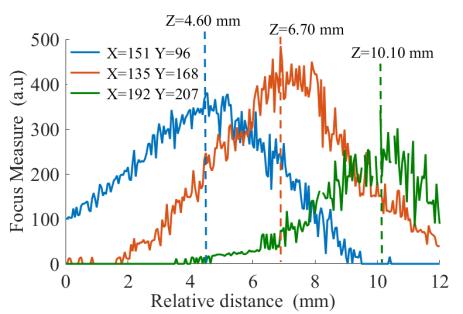
Focus was quantified with a modified Laplacian, an operator that responds to local intensity changes and therefore to edges and fine structure. The calculation used each pixel’s nearest neighbors and accumulated the response over an 11 by 11 window. A 3 dB threshold rejected regions whose contrast was too weak to support a stable depth estimate. The resulting focus metric was evaluated along the axial stack and fitted with a Gaussian curve. The peak position supplied the local depth; the image value at or near that peak supplied the focused intensity.
A second experiment made the problem more demanding by placing a maple leaf over the oak leaf with a separation that varied from roughly 10 to 20 mm. This acquisition contained 1,292 images. A pixel could now produce two focus peaks, one for each layer. The authors applied a second-order Butterworth low-pass filter with a 1 mm-1 cutoff before interpolating the peaks. The filtering reduced fluctuations while preserving the two maxima associated with the separated leaves.
Depth maps, layered samples and practical limits
For the single oak leaf, the reconstructed all-in-focus image recovered veins that were blurred in individual frames. The corresponding depth map followed the leaf’s curved surface. For the two-leaf assembly, the analysis separated the maple and oak surfaces and reconstructed their different shapes despite attenuation through the upper layer. These are controlled demonstrations on thin natural samples, but they show that full-field terahertzTerahertz radiation is electromagnetic energy commonly associated with frequencies around 0.1 to 10 THz, between microwaves and infrared, where many materials reveal distinctive propagation, absorption, and imaging behavior. More data can support rapid surface reconstruction without adding interferometric or tomographic hardware.
The reported lateral resolution was about 0.35 mm, while depth discrimination was limited to approximately 3 mm by the Rayleigh distance. Two surfaces closer than that produce overlapping focus curves that cannot be separated reliably with this configuration. Dead pixels and regions with poor signal-to-noise ratio also leave holes or unstable points in the map. Changing the optics could reduce the depth of field and sharpen axial discrimination, but that choice affects field of view, light collection and tolerance to sample position.
Shape-from-focus also depends on visible texture. A perfectly uniform region offers little Laplacian response even when it is geometrically in focus. Strong absorption can hide a lower layer, and a depth peak identifies the position of maximum focus rather than the material composition of the surface. The method should therefore be viewed as a geometrical reconstruction tool that complements, rather than replaces, spectroscopic identification or computed tomography.
The collaboration combines terahertzTerahertz radiation is electromagnetic energy commonly associated with frequencies around 0.1 to 10 THz, between microwaves and infrared, where many materials reveal distinctive propagation, absorption, and imaging behavior. More source and camera engineering with image-processing expertise. Its practical result is an acquisition-and-analysis route that uses existing full-field hardware and requires no added optical component. For non-destructive testingInspection of dielectric, layered, polymer, composite, and manufactured structures without destructive sampling. Non-Destructive Testing: measurement approach and use cases Work begins with the material and the decision that the measurement... More, it could support rapid measurement of thin formed parts, separated layers or surfaces whose curvature makes one fixed focal plane inadequate. Transfer to a production setting would still require material-specific contrast tests, calibration of axial motion, treatment of vibration and dead pixels, and verification that acquisition speed remains compatible with the required depth precision.
The study’s central insight is economical: depth information can be recovered from the same defocus that ordinarily reduces image quality. Under the tested conditions, a ten-second stack was sufficient to turn a sequence of partially blurred frames into a focused terahertzTerahertz radiation is electromagnetic energy commonly associated with frequencies around 0.1 to 10 THz, between microwaves and infrared, where many materials reveal distinctive propagation, absorption, and imaging behavior. More image and a three-dimensional surface map. That establishes a useful laboratory capability while leaving clear engineering choices around optical depth of field, detector quality and the trade-off between inspected area and axial resolution.
Publication and citation
Recommended citation: Perraud, J.-B., Guillet, J.-P., Redon, O., Hamdi, M., Simoens, F., & Mounaix, P. (2019). Shape-from-focus for real-time terahertzTerahertz radiation is electromagnetic energy commonly associated with frequencies around 0.1 to 10 THz, between microwaves and infrared, where many materials reveal distinctive propagation, absorption, and imaging behavior. More 3D imaging. Optics Letters, 44(3), 483. https://doi.org/10.1364/OL.44.000483
Publisher: Optica Publishing Group. Airtable record: recTQvFrGlEXqtuuS.