A three-dimensional terahertzTerahertz radiation is electromagnetic energy commonly associated with frequencies around 0.1 to 10 THz, between microwaves and infrared, where many materials reveal distinctive propagation, absorption, and imaging behavior. More scan is useful only when its reconstructed voxels can be turned into trustworthy information about an object. This paper presents an end-to-end processing sequence that moves from continuous-wave projection data to segmented volumes, centerline geometry and local dimensional measurements. The contribution is therefore broader than a reconstruction algorithm: it is a workflow for converting terahertzTerahertz radiation is electromagnetic energy commonly associated with frequencies around 0.1 to 10 THz, between microwaves and infrared, where many materials reveal distinctive propagation, absorption, and imaging behavior. More attenuation data into quantities that can support non-destructive inspection.
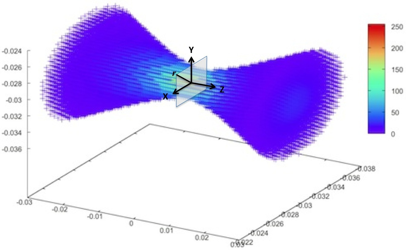
Featured visual: Contextual research figure from âOrdered subsets convex algorithm for 3D terahertzTerahertz radiation is electromagnetic energy commonly associated with frequencies around 0.1 to 10 THz, between microwaves and infrared, where many materials reveal distinctive propagation, absorption, and imaging behavior. More transmission tomographyâ. It illustrates a closely related terahertzTerahertz radiation is electromagnetic energy commonly associated with frequencies around 0.1 to 10 THz, between microwaves and infrared, where many materials reveal distinctive propagation, absorption, and imaging behavior. More topic and is not a figure from the publication discussed on this page. Source publication.
Visuals are drawn from the Airtable research archive. Figure numbering, rights and interpretation should be checked against the original publication before republication outside this site.
From projection acquisition to a regularized THz volume
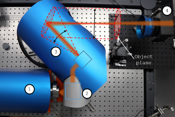
The scanner used a Gunn diode followed by frequency multiplication to generate 287 GHz radiation with a reported power of 12 mW. PTFE lenses collimated and focused the beam onto samples mounted on a motorized translation-and-rotation stage. A zero-bias Schottky diode measured the transmitted signal, with 1 kHz modulation and lock-in detection. Projections were collected over angles spanning 0 to 180 degrees. Depending on the object, the spatial sampling could reach 0.5 mm, while individual projection times and complete scans remained on the scale of minutes and hours rather than real-time acquisition.
The reconstruction stage used a regularized form of the TerahertzTerahertz radiation is electromagnetic energy commonly associated with frequencies around 0.1 to 10 THz, between microwaves and infrared, where many materials reveal distinctive propagation, absorption, and imaging behavior. More Ordered Subsets Convex algorithm. Its forward model includes the Gaussian beam profile, which is essential because the 287 GHz field is not a perfectly narrow ray and changes as it propagates through a three-dimensional object. Iterative updates estimate the attenuation assigned to each voxel. A regularization term suppresses weak fluctuations inside otherwise homogeneous regions while retaining stronger boundaries. The output is a volumetric attenuation map, not yet a finished engineering measurement.
This distinction motivates the rest of the processing sequence. Raw or reconstructed intensity alone does not identify which voxels belong to the component, whether two regions are connected, or how a curved internal channel changes in diameter. Those questions require additional image analysis, especially when background noise and low-contrast material responses occupy overlapping numerical ranges.
Segmentation, topology and local dimensions
The authors first applied K-means clustering to the voxel intensities. For the demonstrated objects, three clusters offered a useful separation: one low-intensity class represented air and background, while two higher-intensity classes captured parts of the object with different attenuation. Combining the object classes produced a binary volume without requiring a manually selected global threshold. Connected-component labeling then separated distinct volumes of interest so that mechanically or spatially independent parts could be analyzed on their own.
Each selected volume was reduced to a one-voxel-thick skeleton through topology-preserving thinning. The skeleton represents the medial organization of a shape as branches and junctions. For a tubular part, it provides a natural path along which dimensions can be sampled. The caliber procedure constructs a plane approximately normal to the local skeleton direction, intersects that plane with the segmented boundary and measures distances across the resulting section. Repeating this operation along a branch yields profiles of outside diameter, internal diameter or wall thickness, together with summary statistics.
The pipeline was demonstrated on three objects with different geometric challenges: a correction-tape roller, a cross-shaped tube and a manual propipetter with a bulb and valves. The segmented volumes enabled surface and volume calculations, while skeletonization exposed the organization of channels and subcomponents. For a straight branch of the cross-tube, the reported mean lumen diameter was about 4.3 mm and the wall thickness about 0.6 mm, close to direct measurements within the resolution limits of the scan. The propipetter example showed how the same approach could follow a more complex pathway and locate changes around valve chambers.
Value and limitations for non-destructive evaluation
The main practical advance is the continuity of the workflow. TerahertzTerahertz radiation is electromagnetic energy commonly associated with frequencies around 0.1 to 10 THz, between microwaves and infrared, where many materials reveal distinctive propagation, absorption, and imaging behavior. More tomography had already demonstrated the ability to image low-loss polymers and related materials, but this sequence connects acquisition to explicit geometric descriptors. Such measurements are relevant to internal channels, polymer housings, lightweight assemblies and manufactured components that cannot be inspected optically once assembled. They can also guide comparison between a measured object and its nominal geometry.
The results remain bounded by the source wavelength, beam waist, detector response and reconstruction assumptions. At 287 GHz, the beam diameter is on the millimeter scale, so very thin walls or closely spaced details may merge. Refraction, scattering and interfaces can change the signal in ways not fully represented by attenuation alone. K-means also depends on the distribution of voxel values; a more heterogeneous, multi-material object may require more adaptive classification or measurements at several frequencies. Skeletons extracted from noisy boundaries can acquire false branches and therefore need quality control before dimensions are accepted.
The study is a clear example of collaboration between terahertzTerahertz radiation is electromagnetic energy commonly associated with frequencies around 0.1 to 10 THz, between microwaves and infrared, where many materials reveal distinctive propagation, absorption, and imaging behavior. More hardware and computational morphology. The author and publication metadata support that interdisciplinary reading, but they do not establish a certified industrial metrology process. A future qualification route would require traceable phantoms, uncertainty budgets, repeatability tests and faster acquisition. Multispectral data could additionally separate materials rather than relying only on attenuation levels.
Even with those caveats, the paper shifts the purpose of three-dimensional THz imaging from seeing an internal form to measuring it. By combining physics-aware reconstruction with segmentation and centerline-based analysis, it provides a reusable framework for extracting dimensions from volumes where conventional visual inspection cannot reach.
Publication details and citation
Recommended citation: Balacey, H., Recur, B., Perraud, J.-B., Bou Sleiman, J., Guillet, J.-P., & Mounaix, P. (2016). Advanced Processing Sequence for 3-D THz Imaging. IEEE Transactions on TerahertzTerahertz radiation is electromagnetic energy commonly associated with frequencies around 0.1 to 10 THz, between microwaves and infrared, where many materials reveal distinctive propagation, absorption, and imaging behavior. More Science and Technology, 6(2), 191-198. https://doi.org/10.1109/TTHZ.2016.2519263
Record ID: recKSg3kwSxMLi8dE
Research themes: 3D terahertzTerahertz radiation is electromagnetic energy commonly associated with frequencies around 0.1 to 10 THz, between microwaves and infrared, where many materials reveal distinctive propagation, absorption, and imaging behavior. More imaging, iterative reconstruction, segmentation, skeletonization, dimensional analysis, non-destructive testingInspection of dielectric, layered, polymer, composite, and manufactured structures without destructive sampling. Non-Destructive Testing: measurement approach and use cases Work begins with the material and the decision that the measurement... More.